The Department of Developmental Physiology‘s primary research focus is the preimplantation developmental period, which encompasses the interval between fertilisation of the egg in the fallopian tube and implantation of the embryo in the uterus. This period is one of the most vulnerable phases in mammalian embryonic development. Disturbances that occur during this period are often the cause of unsuccessful pregnancies in both humans and animals. The research team is currently engaged in investigating the interactions between the maternal organism and the preimplantation embryo and in reparatory mechanisms occurring in early embryos. We examine these processes mainly in the context of the mother’s physiological or health status, such as obesity, stress, or intoxication, using primarily a laboratory mouse model.
- Using a restraint stress model, we demonstrated the harmful effects of maternal stress on early embryonic development and identified embryonic cell receptors involved.
- Using a two-generation diet-induced obesity model, we found that maternal obesity can adversely affect fertilization and preimplantation development, although a certain amount of body fat is necessary to maintain optimal reproductive health.
- We have also demonstrated the negative effects of certain types of insecticides and food additives on early embryo development and identified embryonic cell receptors that may be involved in mediating these effects.
Furthermore, results of our research indicate that maternal stress, obesity or intoxication during early pregnancy can affect the somatic and behavioural parameters of offspring. As part of our study of repair mechanisms in early embryos, we analyzed the occurrence of spontaneous apoptosis in preimplantation embryos of several mammalian species.
In follow-up research, we demonstrated that cells of preimplantation embryos possess all the necessary mechanisms for recognising, engulfing and digesting apoptotic cells, ensuring the clearance of most dying blastomeres. Our latest research focuses on the possibility of preventing early developmental disorders by adding specific additives to the mother’s diet.
The department is comprised of one laboratory:
- Laboratory of Reproduction, Embryology and Cell Signalling

Lab equipment
- Leica TCS SP5 confocal microscope with “live-cell imaging” system for studying the functional morphology of living cells in real time
- Automated Nikon Eclipse 90i direct fluorescence microscope for studying functional cell morphology and morphological diagnosis of microflora
- Olympus BX51 direct fluorescence microscope for the study and functional morphology of cells and morphological diagnosis of microflora (together with NbU)
- Nikon Ti-E inverted fluorescence microscope with micromanipulation kit (Eppendorf) for micromanipulation interventions at the cellular level
- Nikon Ti-U inverted light microscope for studying the morphology of cells cultured in vitro
- DXR Raman spectrometer with microscope (Thermo Fisher Scientific) for metabolomics analysis of cells and biological fluids
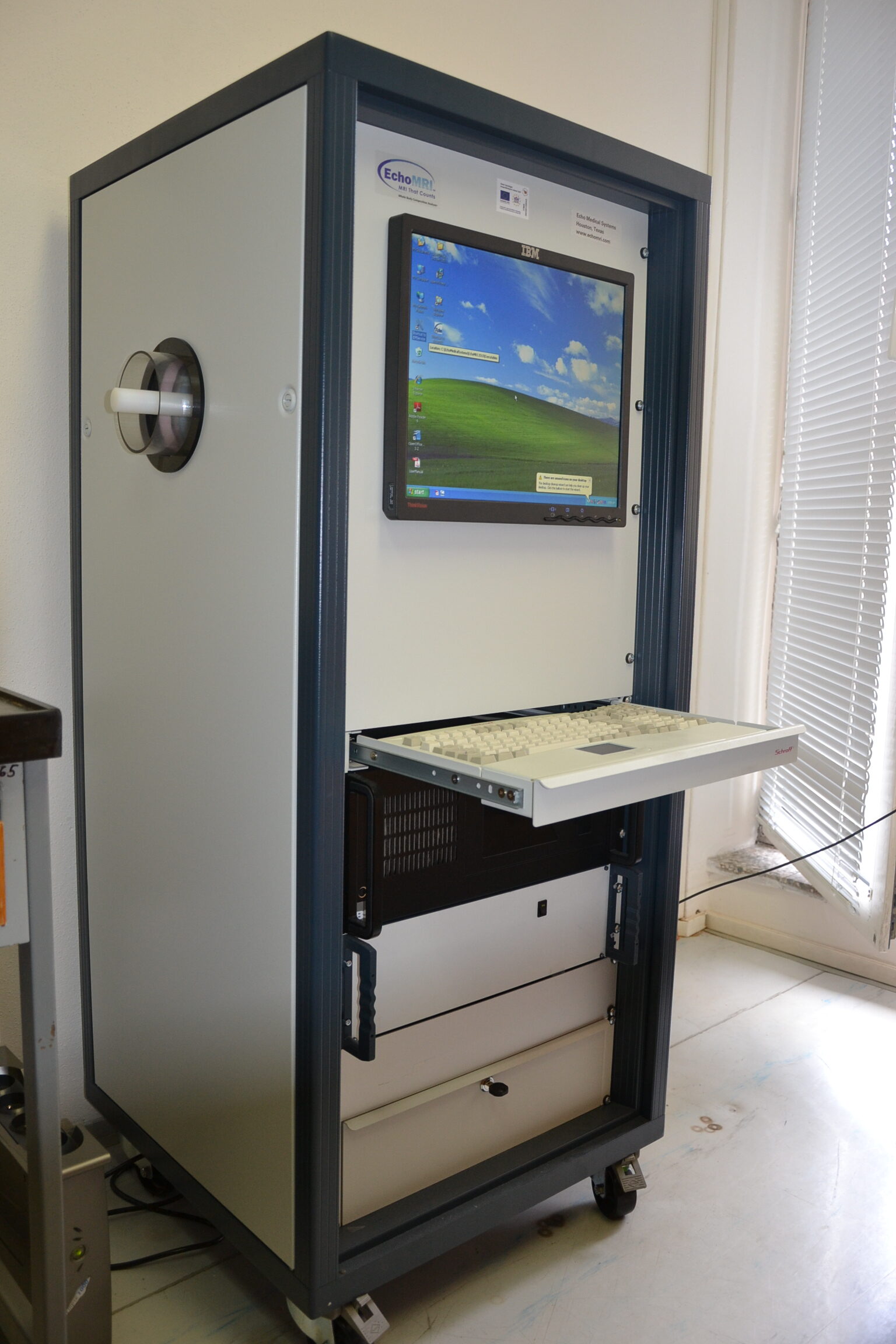
- Whole-body magnetic resonance analyser for laboratory animals EchoMRI 700 for measuring body fat in laboratory animals
- CODA non-invasive blood pressure monitor for laboratory animals
- Real-time PCR system LC480 (Roche) and CFDX Opus (BioRad) for detection and quantification of selected nucleic acid (gene) sequences
- Synergy 2 microplate multidetection analyzer (Biotek) for analyses requiring fluorescence, luminescence and absorbance measurements
- Agilent 2100 capillary microelectrophoretic system for automated electrophoretic analysis of DNA, RNA and proteins.
- Eppendorf multiporator for electroporation of eukaryotic cells, bacteria and yeast
- Fusion FX7 high sensitivity detection imaging system (Vilber Lourmat) for detection of chemiluminescence signal on membranes (Western blot analysis)
- NanoDrop microspectrophotometer (Thermo Scientific) for spectrometric measurement of limited volumes (1 ul) of nucleic acids